
Every emergency clinician remembers the first time a focused ultrasound exam changed a case in real time, free fluid found where none was expected, a pneumothorax confirmed in seconds instead of waiting on a full radiographic study, a rising fluid score that quietly answered whether a patient needed surgery before they crashed. AFAST and TFAST (abdominal and thoracic focused assessment with sonography) were built for exactly this: fast, standardized, repeatable answers to specific clinical questions, performed as an extension of the physical exam rather than a full diagnostic ultrasound study.
The equipment to run these exams has become far more accessible to general practice over the last several years. The harder part, and the part that actually determines whether a clinic gets real value from having a machine, is building genuine team proficiency with the actual protocol, not a loose approximation of it.
Equipment: what you actually need, and what you don’t
One of the more reassuring things about AFAST/TFAST is how modest the equipment requirement actually is. The standard recommendation is a microconvex curvilinear probe, with a frequency range of roughly 5–10 MHz, using the machine’s standard abdominal preset at a depth range of about 3–15 cm, and that single probe and preset combination is generally sufficient for the entire Global FAST exam (AFAST, TFAST, and Vet BLUE together) in most dogs and cats. A phased-array cardiac probe or a linear probe can add value for more detailed follow-up studies, but neither is necessary for the standard screening exam. Adding them mainly adds time without adding diagnostic yield for the triage question you’re actually asking.

This is a good example of where the equipment spec and the actual protocol line up cleanly. The Mindray Vetus E7, for instance, includes a micro-convex probe purpose-built for abdominal scanningand basic cardiology in cats, small dogs, and exotic animals, exactly the transducer profile the AFAST/TFAST protocol calls for, without needing to invest in a separate specialty cardiac probe just to run a standard screening exam. It also ships with dedicated veterinary presets subdivided by species and weight, which matters more than it might sound for a protocol built around consistency. A preset that’s actually tuned to a 4kg cat versus a 40kg dog reduces one more source of variability between operators and between scans.
Worth calling out specifically for the proficiency angle this whole article is about: the Vetus E7 also includes a built-in scanning guidance tool (Mindray calls it iScanHelper) that walks a less experienced operator through how and what to scan for a given exam type. That’s not a substitute for real AFAST/TFAST training, nothing replaces following the standardized protocol properly, but it’s a genuinely useful bridge for a newer team member’s first few dozen scans, while the muscle memory and pattern recognition described below are still being built.
It’s also part of why more veterinary ultrasound training courses have become available specifically built around this exact equipment profile, the protocol was designed from the outset to be teachable and repeatable with accessible, portable gear, not gatekept behind specialist-only equipment.
The other operational principle worth internalizing early: bring the machine to the patient, not the patient to the machine. A portable, cage-side-capable system, under 7 lbs and 1.7 inches thick in the Vetus E7’s case, removes the friction that otherwise discourages a team from reaching for focused ultrasound as often as the clinical picture actually warrants.

The four AFAST windows, specifically
AFAST is not an open-ended abdominal scan. It’s four standardized views, performed in a fixed counter-clockwise sequence, each named for the anatomy it targets:
- DH, diaphragmatico-hepatic view. Probe placed at the subxiphoid space, angled cranially. This view also serves double duty for TFAST, since it images the pericardial and pleural space from the same window, and it’s specifically validated for detecting pericardial effusion, not just abdominal fluid. A 2016 study using the DH view alone identified pericardial effusion in 24 dogs, underlining that this single window is doing more diagnostic work than its “abdominal” name might suggest.
- SR, splenorenal view. Probe placed at the left flank, imaging the spleen and left kidney, also useful as a window into the retroperitoneal space.
- CC, cystocolic view. Probe on midline over the urinary bladder, imaging the bladder and descending colon.
- HR, hepatorenal view. Probe at the right flank/umbilical region, imaging the liver and right kidney. In higher-scoring patients, this site is often the most favorable location for abdominocentesis if fluid is present.
At each site, the technique is consistent: the probe is moved a few inches in several directions and fanned through roughly a 45-degree angle until the target organs are clearly identified. This is a small systematic search at each window, not a single static image. The exam is traditionally performed in right lateral recumbency, which Dr. Lisciandro’s group found preferable to left lateral because it’s more amenable to simultaneous echocardiography, ECG, gallbladder and caudal vena cava imaging, and abdominocentesis if the exam is positive, though AFAST can also be performed with the patient standing, which is lower-impact for painful or fractious patients who may not tolerate lateral restraint well.
The AFS: what the score actually means
The Abdominal Fluid Score (AFS) is what turns a positive AFAST from a yes/no finding into something you can track over time. It’s a simple 0–4 scale based on how many of the four windows are positive for free fluid, not fluid volume directly, but the number of positive sites:
- AFS 0, no positive sites
- AFS 1, positive at any single site
- AFS 2, positive at any two sites
- AFS 3, positive at any three sites
- AFS 4, positive at all four sites (maximum score)
This matters clinically, not just academically. In the original validation study, 101 dogs with motor vehicle trauma, dogs with an AFS of 3 or 4 showed significantly more pronounced drops in PCV and total protein, higher ALT elevations, and needed more blood transfusions than lower-scoring dogs. In other words, the number of positive windows correlates with how much bleeding is actually happening, which gives you a genuinely useful triage signal beyond “there’s some fluid.”
The AFS is also meant to be repeated, not just measured once. Standard practice is to reassess at four hours in a stable patient, sooner if the patient is unstable, since a rising score over that interval suggests ongoing intra-abdominal hemorrhage, while a falling score is consistent with resolution (most traumatic hemoabdomen cases resolve within about 48 hours once bleeding has stopped). That serial comparison is often more clinically useful than either scan on its own, and it’s the kind of longitudinal data point a physical exam alone simply can’t give you.
TFAST: pneumothorax detection via the glide sign
TFAST answers a different but equally time-sensitive question for the thorax: is there pneumothorax, pleural effusion, or pericardial effusion present? It uses five points, bilateral chest tube site (CTS) views, bilateral pericardial site (PCS) views, and the DH view shared with AFAST for pleural effusion.
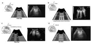
Adapted from Lisciandro et al. (2008), Journal of Veterinary Emergency and Critical Care / Wiley Online Library.
The CTS view is where pneumothorax detection happens, and it hinges on a specific finding called the glide sign: the normal, rhythmic to-and-fro motion of the visceral pleura sliding against the parietal pleura, visible as a shimmering line at the pleural interface. A present glide sign essentially rules out pneumothorax at that location, since air between the pleural layers eliminates that sliding motion. When the glide sign is absent, the next step is searching for the lung point, the specific spot where aerated, sliding lung transitions to the static, air-filled space of a pneumothorax. Where that lung point falls, moving the probe sequentially from dorsal to ventral, helps distinguish a partial pneumothorax (lung point found) from a massive one (no lung point identified at any position, because the lung has collapsed away from the chest wall entirely).
For the pericardial site views, following the standardized probe maneuvers matters more than it might seem. The left PCS view specifically involves sliding the probe cranial and caudal to the heart into two named pouches, the cardiac-diaphragmatic and cardiac-cervical pouches. Following this standardized approach, rather than an improvised look around the heart, is part of what the protocol’s published “TFAST rules” are built to prevent: specifically, misinterpreting normal cardiac chambers as effusion, which is one of the more common interpretation errors in less experienced hands.
This is genuinely high-value information delivered fast: TFAST’s sensitivity for pneumothorax meaningfully exceeds thoracic auscultation alone, and it performs particularly well in patients breathing slowly and deeply, which, notably, is often exactly the patient you’re most worried about.
More than trauma: triage and tracking
It’s easy to think of AFAST/TFAST as a trauma tool exclusively (“for the hit-by-car dog”), but that undersells what the protocol was actually built for. The original framework explicitly covers three clinical contexts, sometimes referred to as T3: Trauma, Triage (any acute, non-traumatic presentation), and Tracking (serial monitoring of hospitalized or critical patients).
In practice, that means AFAST/TFAST has genuine value well beyond the trauma bay:
- Acute collapse or syncope of unknown cause, screening quickly for pericardial effusion, free abdominal fluid, or pneumothorax as part of the initial workup, before deciding what further diagnostics are warranted.
- Suspected GDV or acute abdomen, a fast screen that can inform triage priority and help decide how urgently additional imaging or surgery is needed.
- Anaphylaxis and other acute non-traumatic presentations, where free fluid or effusion may be present without an obvious traumatic mechanism.
- Serial monitoring of hospitalized or post-surgical patients, using repeated AFS or TFAST checks as an objective, trackable data point alongside vitals, rather than relying on physical exam alone to catch a developing problem.
This broader framing matters for a general practice specifically, because it means the return on investment for building real proficiency isn’t limited to the occasional trauma case, it’s a tool that pays off across a much wider slice of daily caseload, especially for tracking hospitalized patients where a change in AFS or TFAST findings can flag a deteriorating patient before vitals alone would.
Global FAST, and the bias it’s specifically designed to prevent
When AFAST, TFAST, and Vet BLUE (the lung-focused component) are performed together as a single, standardized 15-view survey, the combination is called Global FAST. There’s a specific, well-articulated reason for doing all three together rather than just scanning “the area you’re worried about”: it guards against satisfaction of search error and confirmation bias, the well-documented tendency to stop looking once you’ve found something that confirms what you already suspected, potentially missing a second, unrelated finding elsewhere.
A patient presenting after trauma with an obviously painful limb can also have a clinically silent pneumothorax or pericardial effusion that a narrowly focused exam would never catch, simply because nobody was looking there. Performing the full standardized survey as a baseline, rather than a targeted “just check the belly” look, is what protects against that blind spot. It’s also worth being precise about terminology here: Dr. Lisciandro is explicit that Global FAST should never be conflated with a “Flash exam”, a quick, informal look isn’t the same discipline as the standardized, repeatable protocol described above, and the two shouldn’t be used interchangeably even though they can look superficially similar to an untrained observer.
Why proficiency, not just access, is the real bottleneck
A clinic can own an excellent ultrasound system and still get inconsistent value from it if the protocol itself isn’t followed consistently, or if only one team member can run it confidently under pressure. The clinics that get the most value from point-of-care ultrasound are the ones where multiple team members can perform the standardized four-window AFAST and five-point TFAST reliably, not an improvised scan that varies operator to operator.
A few things that genuinely move the needle on team proficiency:
Structured practice on stable patients, not just emergencies. Waiting for a true emergency to be the only time AFAST/TFAST gets practiced means the team’s first real reps happen under the worst possible conditions. Running the four AFAST windows on stable, cooperative patients during routine visits, even with no clinical question to answer, builds the speed and pattern recognition that make the exam fast and reliable when it actually matters, and it gives the team a mental library of what normal looks like at each window, which is what makes an abnormal finding jump out later.
Following the counter-clockwise sequence the same way every time. Proficiency comes from repetition of a standardized approach, same window order, same probe orientation, same fan technique at each site, not from treating every scan as a fresh improvisation. This is also what makes the AFS meaningful: the score only means something if the same four sites are checked the same way every time, which is precisely the discipline that separates Global FAST from a “Flash exam.”
Formal CE investment for at least one team champion. Having one team member complete structured, RACE-approved AFAST/TFAST veterinary ultrasound training gives the rest of the team an in-house resource for troubleshooting and mentorship, rather than everyone learning informally from incomplete secondhand knowledge. FASTVet, founded by Dr. Lisciandro himself, is the most direct source for this, both the original published protocol detail and structured online or in-person courses. For practices specifically looking for veterinary ultrasound courses in Canada, it’s worth checking whether a RACE-approved online option covers your continuing education requirements before committing a team member’s time to in-person training abroad.
Equipment that supports cage-side use, not just a dedicated ultrasound room. A lightweight, genuinely portable system, something in the range of the Mindray Vetus E7 or the M9Vet, removes the friction of “the ultrasound is busy in another room” that quietly discourages teams from using focused ultrasound as often as they otherwise would.

Building this into daily practice, not a one-time training event
The teams that get real, lasting value from AFAST/TFAST treat it the same way they’d treat any other core clinical skill, practiced regularly, reviewed periodically, and built into standard triage rather than reserved for the most dramatic cases.
- Add AFAST to your standard trauma/collapse triage protocol as a default step, the same way you’d default to checking a blood pressure or a lactate, not an optional add-on reserved for the clearest cases.
- Use the AFS consistently, and repeat it. A single AFAST tells you what’s happening right now; a repeated AFS at four hours tells you whether the situation is improving or getting worse, and that trend is often the more clinically actionable piece of information.
- Think beyond trauma. Build AFAST/TFAST into your approach to acute collapse workups, suspected GDV triage, and serial monitoring of hospitalized patients, not just the obvious trauma presentation.
- Debrief scans as a team, even informal ones. A quick “here’s what I saw at each window and why” after a scan builds shared understanding faster than solo practice ever will.
- Revisit training periodically. Skills that aren’t used regularly fade, a brief annual refresher, even informal, keeps the whole team’s baseline competency higher than a single training session years earlier.
Bringing it together
AFAST and TFAST have earned their place as standard-of-care triage tools in emergency and critical care medicine, and general practice is increasingly following that lead, not because the equipment got better (though it has), but because a standardized, repeatable protocol with a real scoring system genuinely changes how confidently and how fast you can answer urgent clinical questions, across a far wider range of cases than trauma alone.
The clinics getting the most value aren’t necessarily the ones with the newest ultrasound system, they’re the ones that treated protocol fidelity and team proficiency as seriously as the purchase decision itself. If you’re evaluating whether your team has the right equipment to support this kind of routine, cage-side use, whether that’s your first system or a second, more portable unit dedicated to triage, we’re happy to talk through what would genuinely fit your caseload.
Comparing specific systems for portability and team use? Our side-by-side look at the Mindray Vetus E7 vs. DP-50 is a good next read if you’re actively choosing between options.
Request a Quote →
Frequently Asked Questions
Question: What are the four AFAST windows?
Answer: The diaphragmatico-hepatic (DH), splenorenal (SR), cystocolic (CC), and hepatorenal (HR) views, performed in a fixed counter-clockwise sequence with the patient typically in right lateral recumbency.
Question: What does an AFS (Abdominal Fluid Score) of 3 or 4 mean clinically?
Answer: A higher AFS, based on the number of positive windows out of four, correlates with more significant intra-abdominal hemorrhage. In the original validation study, dogs with an AFS of 3–4 showed more pronounced drops in PCV and total protein and required more blood transfusions than lower-scoring dogs.
Question: How does TFAST detect pneumothorax?
Answer: By assessing the glide sign, the normal sliding motion between the visceral and parietal pleura, at the chest tube site view. A present glide sign rules out pneumothorax at that site; an absent glide sign prompts a search for the lung point, which helps determine whether the pneumothorax is partial or massive.
Question: Is AFAST/TFAST only useful for trauma cases?
Answer: No, the original framework covers Trauma, Triage (non-traumatic acute presentations), and Tracking (serial monitoring of hospitalized patients). It’s used for acute collapse workups, suspected GDV, anaphylaxis, and monitoring critical patients, not only trauma.
Question: What ultrasound probe is needed for AFAST/TFAST?
Answer: A microconvex curvilinear probe with a 5–10 MHz frequency range and the machine’s standard abdominal preset is sufficient for the full Global FAST exam in most dogs and cats, a specialty cardiac or linear probe isn’t required for standard screening.